


New technique provides novel approach to diagnosing ciliopathies
|
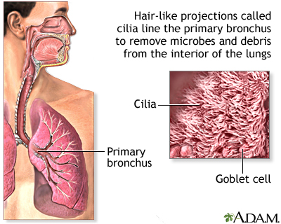
Cilia, the cell’s tails and antennas, are among the most important biological structures. They line our windpipe and sweep away all the junk we inhale; they help us see, smell and reproduce. When a mutation disrupts the function or structure of cilia, the effects on the human body are devastating and sometimes lethal.
The challenge in diagnosing, studying and treating these genetic disorders, called ciliopathies, is the small size of cilia—about 500-times thinner than a piece of paper. It’s been difficult to examine them in molecular detail until now.
Professor Daniela Nicastro and postdoctoral fellow Jianfeng Lin have captured the highest-resolution images of human cilia ever, using a new approach developed jointly with Lawrence Ostrowski and Michael Knowles from the University of North Carolina School of Medicine. They reported on the approach in a recent issue of Nature Communications.
About 20 different ciliopathies have been identified so far, including primary ciliary dyskinesia (PCD) and polycystic kidney disease (PKD), two of the most common ciliopathies. They are typically diagnosed through genetic screening and examination of a patient’s cilia under a conventional electron microscope.
The problem is, conventional electron microscopy is not powerful enough to detect all anomalies in the cilia, even when genetic mutations are present. As a result, the cause of ciliary malfunctions can be elusive and patients with ciliopathies can be misdiagnosed or undiagnosed.
Nicastro and her team developed an approach that includes advanced imaging technique that entails rapidly freezing human samples to preserve their native structure, imaging them with transmission electron microscopy, and turning those images into 3D models. This cutting-edge imaging was in part made possible by the advanced instrumentation in the Louise Mashal Gabbay Cellular Visualization Facility at Brandeis. It is the first time this approach has been used on human cilia and patient samples.
We have a new window into the structure and defects in human cilia.
“For so long, researchers haven’t been able to see the small defects in human cilia,” Nicastro says. “Now, we can fill in the pieces of the puzzle.”
###
Leah Burrows
.(JavaScript must be enabled to view this email address)
781-736-4027
Brandeis University
Print Version
Tell-a-Friend comments powered by Disqus

